
Investigating the Effect of Dopamine on in Vitro Maturation, Fertilization and Development of Immature Bovine Oocytes
Abstract
Proteins and other nitrogenous substances such as polyamines, catecholamines, and nitric oxide require amino acids for synthesis. The purpose of this study was to see how adding dopamine to either maturation or fertilization medium affected the developmental competence of immature bovine oocytes. In this study, Ovaries from apparently normal reproductive organs of cattle were collected within 30 minutes from slaughter and evisceration of animals. Cumulus oocyte complexes (COCs) were collected by aspiration of medium sized ovarian follicles (4-8 mm). COCs of acceptable quality were selected, washed and incubated in tissue culture media (TCM) 199 supplemented with 10% heat inactivated fetal calf serum, 5 μg/ml luteinizing hormone, 0.5 μg/ ml follicle stimulating hormone and 1 μg/ml estradiol-17β for 20:22 hour at 38.5 C◦ under 5% CO2 in air with 90% humidity. different concentrations of dopamine (20,40 ng) were used. The results were consistent for both maturation and fertilization and there is non-significant increase in maturation and fertilization.
Article Information
- Received
- Accepted
- Published
Academic Editor: Betul APAYDIN YILDIRIM, Department of Biochemistry Faculty of Veterinary Medicine Ataturk University Erzurum/TURKEY
Checked for plagiarism: Yes
Copyright © 2024 Marwa EL-Ad, et al.
 This is an open-access article distributed under the terms of the Creative Commons Attribution License, which permits unrestricted use, distribution, and reproduction in any medium, provided the original author and source are credited.
Corresponding author: Mohammed A Elmetwally, Department of Theriogenology, Mansoura University, Mansoura, 35516, Egypt —
Competing Interests
The authors have declared that no competing interests exist.
Funding
No specific funding statement was provided by the authors.
Data Availability
No data-availability statement was provided by the authors.
Citation:
Introduction
Cattle are an important source of meat and milk in Egypt. They are distributed all over the country, with higher density in the Nile valley and delta and usually found in small holdings along with buffaloes. The Egyptian native cattle, called Baladi, had four breeds, being Domiati, Mariuti, Menufi , and Saidi 1.
The Egyptian cattle populations, especially the Egyptian Menufi and Saidi are possessing high genetic variability. Information about comparison between the groups of Egyptian cattle population in Egypt based on their genetic distance value provide some benefits for the Egyptian researchers to design future conservation and breeding programs as well as enrich the Egyptian cattle genetic resources 2.
The increase in the productive efficiency and quality of animal products from livestock has been possible due to the use of novel reproductive biotechniques 3.one of these technologies is in vitro fertilization (IVF) which is a useful tool in performing the selection and breeding of genetically superior animals which is becoming more distinguished in commercial dairies 6.
Laboratory production of embryos (IVF technology) provides an excellent and cheap source of embryos for carrying basic research on developmental physiology, farm animal breeding and for commercial application of the emerging biotechniques like cloning and transgenesis. In vitro fertilization of mammalian ova has been possible for over 20 years 7. Recently, in vitro fertilization by in vitro capacitated bovine sperm been demonstrated and verified by the birth of at least one live calf after transfer of resulting embryos to recipients 8, 9.
Catecholamine (dopamine, epinephrine and norepinephrine) plays an important role during early development of the conceptus via improvement of the amino acid transporter synthesis 10, 11. Amino acids are essential for synthesis of proteins and other nitrogenous substances such as polyamines, catecholamines, and nitric oxide 10. Dopamine (DA), epinephrine (EP), norepinephrine (NE), and serotonin are members of a group of neurotransmitters called biogenic amines. Dopamine and norepinephrine can play an important role in basic developmental processes such as embryogenesis and morphogenesis, as well as proliferation, differentiation, and migration of cells. Ovine trophectoderm cells secrete NE and DA that may have significant effects during the peri-implantation period of pregnancy 11. In gilts, NE and DA increase in uterine flushings between days 12 and 14 of gestation compared to that in uterine flushing from non-pregnant gilts. However, there is no published evidence regarding EP in uterine flushing from cyclic gilts and ewes. Dopamine is not only a precursor to NE and then EP in the biosynthetic pathway for these neurotransmitters, but also an independent neurotransmitter 12.
Results of several studies suggest that DA is important for embryo– fetal development, particularly in motor and cognitive neurological programming. The first meiotic division resumed in vitro when the immature bovine oocytes released and cultured in maturation medium. The changes of basic maturation conditions thought to have significant effects on oocyte competence as reflected by the morula and blastocyst yield after in vitro fertilization (IVF) 13.
Material and Methods
The study was carried out at Animal Reproduction Research Institute - Giza, 12556 Al Ahram -Giza, 12111 Cairo – Egypt during the period from December 2022 to April 2023. The chemicals in this study will be purchased from Sigma Chemical Co (St. Louis, MO, USA). All experimental protocols have been approved (PhD/90) by the Committee for Research Ethics at the Faculty of Veterinary Medicine Mansoura University, Egypt.
Recovery and classification of immature oocytes
The recovery and classification of the immature oocytes were done according to the previous literature 14, 15, 16. In brief, ovaries from apparently normal reproductive organs of heifers and cattle of unknown age and breeding history were collected within 30 minutes after slaughter and evisceration of animals at the private EL-Bagor abattoir. The ovaries were kept in a thermos flask containing warm normal saline and gentamycin. Then transported to the lab within 1-2 h after slaughtering 17. The ovaries were further washed as soon as we arrived to the lab in warm normal saline to remove the blood, debris and then kept in a water bath at 37°C during oocyte collection. Immature oocytes were collected by aspiration of medium-sized (4- 8 mm) ovarian follicles using an 18-gauge needle attached to a disposable 10 ml syringe. Evenly granulated oocytes surrounded with one-layer cell were selected for further experimental procedures. The selected immature oocytes were firstly washed three times in sterile (PBS) phosphate buffer saline solution. Before COCs selection by at least 1 h, the maturation dish(s) is prepared by putting 100µl maturation media drops in sterile disposable petri dish then covered with mineral oil and incubated at 38.5 C◦ under 5% CO2 and 90% humidity 15.
Using of 18-gauge needle attached to a 10 ml syringe, medium sized follicles (3: 8 mm in diameter) were aspirated and pooled in a 15-ml conical tube 18. After the end of aspiration, the tubes were left 10:15 minutes to allow follicular cells settlement.
After sedimentation, about 5ml of sediment was recovered by dropper pipette and placed in an 80 mm diameter sterile petri dish. COCs were selected using a stereomicroscope and transferred into another dish containing fresh pre-warmed TCM199 14, 16.
Classification of recovered cumulus oocyte complexes
The retrieved COCs were classified according to 19 into 4 grades based on their morphological appearance.
Grade A (good), COCs with six layers of dense compact cumulus cells investment and evenly granular homogenous ooplasm.
Grade B (fair), similar to grade A, but with 2-4 layers cumulus cells.
Grade C (poor), partially or completely denuded oocytes.
Grade D (very poor), characterized by highly scattered cumulus cells and dark irregular ooplasm.
Grade A and Grade B COCs were washed three times in TCM199, followed by incubation under mineral oil in the same medium (10:15 oocytes/100 µl) for 22 h at 38.5 C◦ under 5% of CO2 in air with 90% humidity 20.
Semen
Frozen cattle semen was obtained from ARRI. The semen cryopreservation in brief, cattle spermatozoa were collected by AV, evaluated, good quality semen samples were pooled and extended in egg yolk glycerol Tris- based extender and cryopreserved in ¼ ml French straws, each straw contains 25×106 million sperm cell 21
Preparation of basic media
Preparation of media and stock solution requests a sterile technique with exact and careful weighing of components. The use of a laminar flow cabinet is indispensable to evade any contamination that may alter and spoil the prepared media. An analytical balance with readability of at least 10 μg and accuracy of ± 0.1μg was used. All media were filtered using 0.2 μm (Millipore, USA) syringe filter and incubated for at least 2 h in a humidified atmosphere, 5% CO2 at 38.5° C before culturing the oocytes and spermatozoa.
In vitro maturation medium
The medium used for oocyte IVM was TCM-199 supplemented with 10% heat inactivated FCS, 5 μg/ml LH, 0.5 μg/ml FSH and 1 μg/ml estradiol-17β 22. The pH was adjusted to 7.4.
In vitro fertilization medium
In vitro culture medium
The medium used for culture is Modified synthetic oviduct (MSOF) supplemented with 1 mM glutamine, 1% MEM (Eagle′s Minimum Essential medium) nonessential amino acids, 0.5% MEM essential amino acids and 10% FCS24 . The pH was adjusted to 7.4.
Fixation and Staining agents
Fixative, stain and stain removing agents are prepared according to 25.
Acetic acid- ethanol fixative (1:3 v/v).
Aceto-orcein stain: 1% (w/v) orcein stain in 45% acetic acid. Stain differentiation solution is acetic acid, distal water and glycerol (1:3:1 v/v/v).
Semen preparation and Oocyte insemination
Three straws of frozen semen were thawed for 30 sec in a 37C◦ water bath, evacuated in a test tube. Swim-up technique, in modified-sperm TALP medium, was used for separation of motile sperm 26. Dilution of the final pellet of spermatozoa is made to obtain a sperm concentration of 2 million/ml as a final sperm cell concentration 19. 100 µl drops of sperm-TALP media containing spermatozoa are deposited in a sterile disposable petri dish, and then covered with mineral oil and incubated at 38.5 C◦ under 5% CO2 and 90% humidity.
Mature oocytes were washed three times in sperm-TALP then added to the fertilization drops, 10 oocytes in each drop. Gametes were co-incubated at 38.5 C◦ under 5% of CO2 and 90% humidity for 21 h 19.
Inseminated oocyte culture
The culture dish is prepared by putting 50µl culture media drops in a sterile disposable petri dish then covered with mineral oil and incubated at 38.5 C◦ under 5% of CO2 and 90% humidity. After 20-22 h of gametes co-incubation, presumptive zygotes were removed from fertilization droplets. These zygotes were washed three times in the culture medium. The presumptive zygotes (10 zygote/50 μl droplets) were cultured at 38.5 C◦ under 5% of CO2 and 90% humidity 20. Semi replacement of the culture medium by fresh medium is done every 48 h 24.
Maturation rate assessment
Matured oocytes were fixed and stained with aceto-orcein stain 1%. Maturation was indicated by germinal vesicle breakdown 25.
Fertilization rate assessment
Presumptive zygotes were fixed and stained with aceto-orcein stain 1%. Normal fertilization was indicated by presence of two pronuclei 25.
Cleavage and Blastocyst formation rate assessment
Zygotes were assessed for cleavage 48 h after the beginning of culture and further embryo development was checked approximately every 48 h for up to 8 days in order to confirm the blastocyst rate 27.
Fixation and staining method
Presumptive mature oocytes and fertilized oocytes were fixed and stained according to 25.
Statistical analysis
Each experiment was replicated at least three times. The normality of quantitative parameters was assessed using normal probability plots and the Kolmogorov-Smirnov test generated with the UNIVARIATE procedure of SAS according to Elmetwally et al.2018 and 2019 10, 28. All experimental data are expressed as mean ± SEM. Rate of recovery, cleavage and maturation and blastocyst rates are expressed as percentages. Statistical analyses will be done using SAS® (version 9.2, SAS Institute, Cary, NC, USA). Differences will be considered to be significant at (P ≤ 0.05).
Results
Data presented in Table 1 and Figure 1 demonstrated no significant increase in the maturation rate due to dopamine supplementation of in vitro maturation media.
Table 1. Effect of in vitro maturation media supplementation with dopamine on the maturation rate.| Treatment | No of oocytes | Maturation rate (%) |
| Control | 100 | 70±4.61 a |
| 20ng | 100 | 73±4.46a |
| 40ng | 101 | 73.27±4.43a |
Figure 1. Effect of in vitro maturation media supplementation with dopamine on the maturation rate.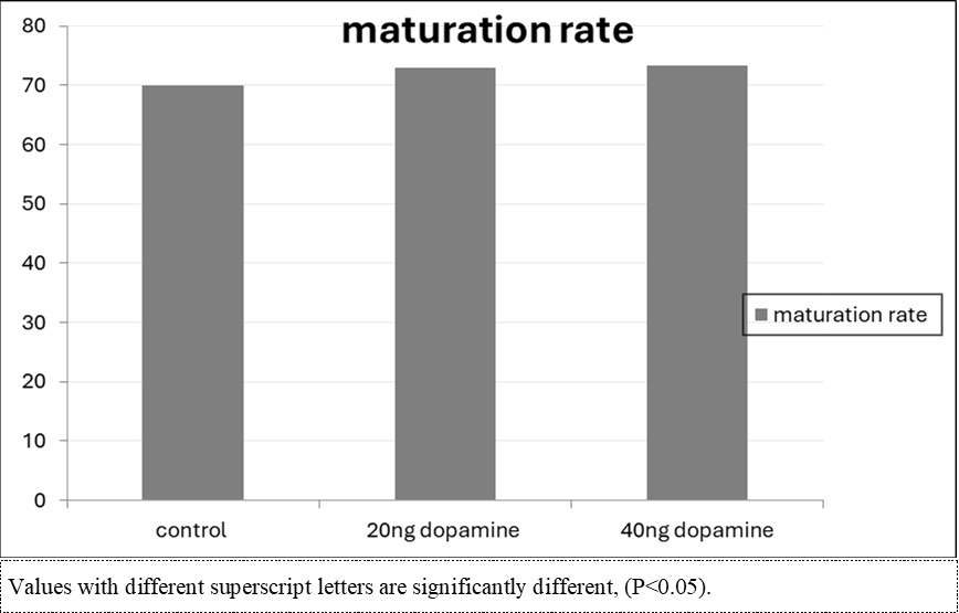
Download figure
Data in Table 2 and Figure 2 indicated that in vitro maturation media supplementation with dopamine led to no significant increase in the fertilization rate.
Table 2. Effect of in vitro maturation media supplementation with dopamine on the expansion rate.| Treatment | No of oocytes | Expansion rate (%) |
| control | 200 | 80.50±2.81a |
| 20ng | 201 | 82.59±2.68a |
| 40ng | 204 | 81.86±2.70a |
Figure 2. Effect of in vitro maturation media supplementation with dopamine on the expansion rate. 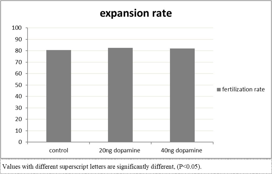
Download figure
As shown in Table 3 and Figure 3, in vitro maturation media supplementation with dopamine led to no significant increase in the cleavage and blastocyst formation rates.
Table 3. Effect of in vitro maturation media supplementation with dopamine on the cleavage and blastocyst formation rates.| Treatment | No of oocytes | Cleavage rate (%) | Blastocyst formation rate (%) |
| control | 100 | 40±4.92 | 10±3.12a |
| 20ng | 100 | 43±4.98 | 11±3.14a |
| 40ng | 102 | 42.16±4.91 | 10.78±3.09a |
Figure 3. Effect of in vitro maturation media supplementation with dopamine on the cleavage and blastocyst formation rates.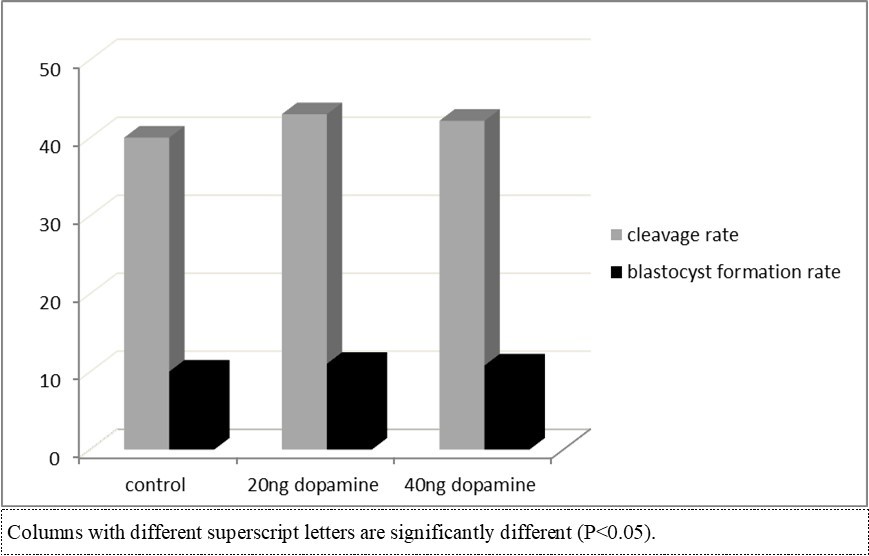
Download figure
Discussion
Catecholamines are tiny, polar, chemical compounds that are produced in neural ectoderm-derived cells from phylogenetically evolved mammals. In the animal kingdom, these compounds serve as related hormones and neurotransmitter substances 29, 30. Other roles for catecholamines and related compounds include those of L-dopa and dopamine in tissue pigmentation and melanin synthesis in insect and mammalian cuticula 31. Apart from these proven roles, there is growing evidence that catecholamines may play a role in cell growth and physiology. This theory is supported by two noteworthy observations. The molecules are found in lower evolutionary forms, such as those seen in protozoans, which are unicellular animals devoid of nerve systems 32, 33, 34.
Dopamine governs several physiological and behavioral processes, including reproduction, which is mediated by the hypothalamic-pituitary-gonadal (HPG) axis in vertebrates. The release of gonadotropin-releasing hormone (GnRH1) from the hypothalamus induces pituitary gonadotropic cells to release luteinizing hormone (LH) and follicle-stimulating hormone (FSH) into the bloodstream. These gonadotropic hormones affect reproductive capacity directly by stimulating the manufacture of gonadal steroid hormones such as testosterone, estrogen, and progestin 10. The present study aimed to highlight the effects of the dopamine on the developmental competence of buffalos’ oocytes. Previous studies by Dr. Elmetwally indicated that not only Dopamine but also other catecholamines have significant effects during the early development window of the fetus 10.
In the present study, the addition of either 20 and/or 40 ng Dopamine to the maturation media increased the maturation rats but the increase was not significant. This may be attributed to the low number of the oocytes or may be regarding the buffalo species. Of note, many scientists have long been fascinated by oocyte maturation and morphogenesis. Despite extensive research on the subject, many parts of the development process remain unknown. One particular area of focus is the interaction of neurotransmitters and neuropeptides with oocyte maturation and early development 35. Due to the limitation of the resources in the present study, we mainly depend on the previous studies that illustrate more biological and molecular studies in addition to Dr. Elmetwally previous studies at Texas A&M University in the USA to build the discussion of the present results. The main function of the dopamine as well as all neurotransmitters depends on Ca2+ signaling machinery as a critical modulator of oocyte physiology and has been thoroughly investigated in a variety of taxa, including mammals 36. Basically, neurotransmitters and neuropeptides including dopamine are connected to Ca2+ during oocyte maturation 37, 38. Although Ca2+ signaling and hormone regulation are thoroughly researched in most phases of early development, there is less evidence on the relationship between neuronal signaling and Ca2+ during important reproductive events 38.
For more information, the neurotransmitters have different signaling pathways, such as fibroblast growth factor (FGF) 39 . Also, Dopamine may play role in Wnt, transforming growth factor- (TGF), and Notch, have been identified as critical regulators of early organism development 39. Chemical or hormonal messengers are typically used to transmit these signals. Several studies, on the other hand, have been successful in demonstrating the role of numerous neurotransmitters and neuropeptides in processes such as oocyte maturation and embryo development 37, 40. Furthermore, the neurotransmitters and neuropeptides have been documented to play a role in oocyte maturation and early development in both vertebrates and invertebrates, as well as present known linkages to Ca2+ signaling throughout these critical processes 37. Although the present results showed no significant differences in the maturation of the immature bovine oocytes.
Also, contrary to the present results dopamine is used broadly in the fish industry; the dopamine is used to prime female broodstocks with various neurotransmitters and neuropeptides. Biogenic amines, two of which are neurotransmitters, serotonin (5-HT) and dopamine (DA), are thought to govern the production and release of neurohormones in crustaceans 41. The results of the previous literature may indicate that the Dopamine may become significantly clear with increasing the oocytes number or we may need to add some other gradient to the maturation media.
Effect of in vitro maturation media supplementation with dopamine on the expansion rate.
In the current study, we highlight for the first time the effects of dopamine on the expansion rate of the immature bovine oocytes. In the present study, all concentrations of the dopamine in the present study showed more than 80 % of immature oocyte expansion. These data indicate that either 20 and/or 40 ng of the dopamine did not have a detrimental effect on the oocyte expansion. Form the reproduction biology point of view, it appears that normal ovulation and subsequent fertilization depend on the cumulus mass expanding to its optimal size 42, 43, 44 found a correlation between the rate of development to the 2-cell stage and the extent of cumulus expansion. Hyaluronic acid (HA) is the main constituent of the enlarged cumulus 45, 46, and the degree of expansion is directly connected to the amount of HA synthesis 36. Research has demonstrated that FSH stimulates the manufacture of HA novo in vitro 47, 48.
The process by which an immature oocyte halted at prophase of the first meiotic division continues meiosis to become competent for regular fertilization following ovulation is known as oocyte maturation 46. Oocytes are initially stopped during the prophase of meiosis I in nearly every species that was studied. Oocytes undergo meiotic maturation when under proper hormonal control, and depending on the species, they halt again at either metaphase I or II. Oocytes are referred to as mature oocytes or eggs at this point, and they will stay in this state until fertilization 49. The oocyte goes through cytoplasmic and nuclear maturation during this transition, which results in the development of the competent egg. Oocyte maturation is recognized to be primarily stimulated by the complex of cyclin B and cyclin-dependent kinase CDK1, known as maturation promoting factor (MPF). Increased cAMP levels prevent MPF activation in stopped oocytes. Germinal vesicle breakdown (GVBD) and chromosomal segregation are initiated when cAMP levels fall, which is accompanied by an increase in MPF activity 50.
The present results regarding the effect of dopamine on the expansion of the immature bovine oocytes are supported by the great evidence that neurotransmitters, neuropeptides and calcium plays a crucial role in oocyte maturation and early development. In general, chemical signals known as neurotransmitters and neuropeptides are found throughout the body and regulate a variety of intricate functions. These chemical signals regulate everything from our mood, sleep patterns, and appetite to autonomic processes like breathing and heart rate. They do this by modifying electrical impulses that are sent from neurons to the body, which changes how the body reacts to various stimuli. Small-molecule chemical messengers called neurotransmitters include glutamate, acetylcholine, histamine, serotonin, and gamma aminobutyric acid (GABA). However, according to their appearance under electron microscopy, neuropeptides—a particular class of large-molecule neurotransmitters —have been identified as dense-core vesicles 51. Neurotransmitter receptors fall into two categories: ionotropic and metabotropic. Ion channels regulated by neurotransmitters are called ionotropic receptors. Ionotropic receptors allow information to be sent between neurons in less than a millisecond. This family of channels opens upon neurotransmitter binding 52. However, because they activate intracellular signaling proteins known as heterotrimeric G proteins, metabotropic receptors are G protein coupled receptors (GPCR), also referred to as seven-transmembrane domain receptors. According to several studies 53, 54, metabotropic receptors are really the mechanism of action for the majority of neuropeptides and small-molecule neurotransmitters.
Effect of in vitro maturation media supplementation with dopamine on the cleavage and blastocyst rate. More than fifty years have passed since the physiological roles of 3-hydroxytyramine, or dopamine, a metabolite of the amino acid tyrosine, were discovered 55. This catecholaminergic neurotransmitter has garnered a great deal of interest. Like other monoamine neurotransmitters, dopamine typically affects neuronal circuitry by modulating rapid neurotransmission, which is mediated by glutamate and GABA, in a relatively slow way. In the brain, dopaminergic innervations are the most prevalent. The mammalian brain is known to contain four major dopaminergic pathways: the nigrostriatal, mesolimbic, mesocortical, and tuberoinfundibular systems. These pathways arise from the dopamine-containing cells in the A9 (nigrostriatal), A10 (mesolimbic and mesocortical, often referred to as the mesocorticolimbic pathway), and A8 (tuberoinfundibular) groups 56, 57, respectively. These neurons play a crucial role in the central nervous system's ability to perform several essential processes, such as working memory, learning, emotion, reward, sleep, and voluntary movement. Dopamine is involved in peripheral physiological processes that include immune system regulation, respiratory function, cardiovascular function, olfactory perception, retinal processes, hormonal regulation, and renal function regulation 58, 59, 60.
In the current study, we also investigated the effects of the dopamine supplementation of maturation media on the cleavage and blastocyst rates of the bovine oocyte. In recent years, new information in the field of assisted reproductive technologies has enabled researchers and practitioners to achieve significant milestones in oocyte and sperm in vitro competence either in bovine 15, 16 or in murine 7. Gamete competence is defined as the capacity to fertilize successfully and produce a normal blastocyst capable of implanting in the uterus and producing healthy offspring 15. Many scientists are working to uncover cellular and molecular indicators that will allow them to pick the most competent oocyte and spermatozoon to produce embryos with higher implantation potential 15.
In the current study for the first time we evaluated the effects of maturation media supplementation with different concentrations of Dopamine on the cleavage and blastocyst formation of immature bovine oocytes. Both developmental parameters showed a non-significant increase with a higher concentration of dopamine. Basically, the precise wiring of the neural circuits is mostly self-generated and depends on the activity of neurotransmitters and neuromodulators, even if genes primarily control the formation of the CNS's scaffold 61, 62. They have the ability to stimulate, intensify, inhibit, block, or lessen the micro-electric signals that are transmitted to neurons 62. As a result, they give rise to the signaling patterns that form the physical networks of brain neurons among countless neural networks. Even before neurons differentiate, neurotransmitters like catecholamine can be seen in the embryos of both vertebrate and invertebrate species 62. Certain neurons in the neural crest are initially noradrenergic, but external stimuli cause them to change to a cholinergic state 35. It was proven that Catecholamine' traditional function is to act as signaling molecules, connecting impulses from a neuron to an effector organ or from another neuron to another. Nevertheless, neurotransmission may play merely one of these molecules' signaling functions. As morphogens in developing embryos, they may have a more rudimentary function 63.
Morphogens are chemicals that have a range of effects on embryonic patterning and gene activity. The evidence for catecholamines' involvement in this process has been compiled in a number of reviews and is based on descriptive examples from a wide range of animal species at different developmental stages 35, 64. For instance, dopamine seems to control the development of its synaptic target neurons in the rodent corpus striatum. These previous literatures indicate that dopamine plays an important role during the early development of the fetus inside the uterus.
In conclusion, dopamine may have a beneficial role in the assisted bovine reproduction. I would like to say that the dopamine as a member from the catecholamines may need further studies to illustrate the present results. Why did dopamine not have a detrimental effect on the immature bovine oocyte? What is the exact concentration of catecholamine in the bovine uterine flush as the present experimental design was mainly based on the proved results published by the reproduction group staff at Dr. Bazer biology lab of pregnancy, Texas A&M, College station, USA, regarding the evidence of presence of catecholamine in the ovine uterine flush. They published for the first time the discovery of epinephrine (Reproduction biologists used to say: epinephrine is the bad guy of catecholamine) beside both norepinephrine and dopamine and their effects on the preimplantation changes as well as its effects on the section of maternal recognition factor. They further studied the effects of dopamine on the expression of polyamines, apoptotic and angiogenic proteins by ovine Trophectoderm cells (oTr1 cell).
References
- 1.MoDAD. (2004) Secondary guidelines for development of national farm animal genetic resources management plans, measurement of domestic animal diversity: Recommended microsatellite markers [Internet]. [cited.
- 2.E M Ghoneim, E F Abbas, A R Elbeltagy. (2018) GENETIC CHARACTERIZATION OF EGYPTIAN CATTLE POPULATIONS IN COMPARISON WITH FRENCH BREEDS. Menoufia Journal of Animal Poultry and Fish Production 2(3), 57-58.
- 3.H A Sharawy, A O Hegab, M A Elmetwally. (2023) Impact of the corpus luteum and follicular parameters on the levels of plasma progesterone and the prediction of pregnancy in Holstein dairy cows. Reprod. Domest Anim 58, 1525-1531.
- 4.H A Sharawy, A O Hegab, E F Risha, El-Adl M, W T Soliman et al. (2022) The vaginal and uterine blood flow changes during the ovsynch program and its impact on the pregnancy rates in Holstein dairy cows. BMC Vet. Res 18, 350.
- 5.Gohar M, Zaabel S, Eldomany W, Eldosouky A, Tawfik W et al. (2023) Transrectal Doppler Ultrasound to Study the Uterine Blood Flow Changes During the Puerperium in the Egyptian Buffaloes. Journal of Advanced Veterinary Research
- 6.M A Sirard. (2018) 40 years of bovine IVF in the new genomic selection context. Reproduction 156(1), R1-R7.
- 7.Helmy A, Halawa A, Montaser A, Zaabel S, Tawfik W et al. (2023) The effects of fructose on the developmental competence, biochemical biomarkers and apoptotic gene expression by murine oocytes. NIDOC-ASRT 54, 323-336.
- 8.B G, Bousquet D, M L Boice, W J Donawick, J F Evans et al. (1982) Normal development following in vitro fertilization in the cow. Biology of reproduction 27(1), 147-158.
- 9.B G, Y K Oh, J F Evans, W J Donawick. (1980) Fertilization and early development of cow ova. Biology of Reproduction 23(1), 189-205.
- 10.M A Elmetwally, Lenis Y, Tang W, Wu G, F W Bazer. (2018) Effects of catecholamines on secretion of interferon tau and expression of genes for synthesis of polyamines and apoptosis by ovine trophectoderm. Biology of Reproduction, 99(3). 611-628.
- 11.Y, M A Elmetwally, Tang W, Satterfield C, Dunlap K et al. (2018) Functional roles ofagmatinaseduring the peri-implantation period of pregnancy in sheep. Amino Acids 50, 293-308.
- 12.Y, M A Elmetwally, J G Maldonado-Estrada, F W Bazer. (2017) Physiological importance of polyamines. Zygote 25(3), 244-255.
- 13.B D, T A Rose-Hellekant, Pinyopummintr T. (1992) Development of in vitro matured/in vitro fertilized bovine embryos into morulae and blastocysts in defined culture media .
- 14.Mostagir A, Elmetwally M, Montaser A, Zaabel S. (2019) . Effects of L Carnitine and Cryoprotectants on Viability Rate of Immature Buffalo Oocytesin vitro After Vitrification. AJVS 62 45.
- 15.M A Elmetwally, A M Mostagir, F W Bazer, Montaser A, Badr M et al. (2022) Effects of L-carnitine andcryodeviceson the vitrification and developmental competence of invitro fertilized buffalo oocytes. , Veterinary Medical Society, Journal of the Hellenic
- 16.Abdeldayem M, Zaabel S, Badr M, Sharawy H, Mostagir A et al. (2022) Effects of L arginine supplementation on in-vitro maturation and cryotolerance of immature buffalo oocyte. Mans. Vet. Med. J 0, 0-0.
- 17.A-s A-H El-Naby, K G Mahmoud, Y F Ahmed, M E Abouel-Roos, A E. (2013) Effect of Season of the year and Ovarian Structures on Oocytes Recovery Rate, Quality and Meiotic Competence in Egyptian Buffaloes. Glob Vet 10, 408-412.
- 18.Raghu H M, Nandi S, Reddy S M. (2002) Follicle size and oocyte diameter in relation to developmental competence of buffalo oocytes in vitro. Reprod Fertil Dev 14, 55-61.
- 19.Neglia G, Gasparrini B, V Caracciolo di Brienza, R Di Palo, Campanile G et al. (2003) Bovine and buffalo in vitro embryo production using oocytes derived from abattoir ovaries or collected by transvaginal follicle aspiration. Theriogenology 59, 1123-1130.
- 20.Gasparrini B, A De Rosa, Attanasio L, Boccia L, R DI Palo et al. (2008) Influence of the duration of in vitro maturation and gamete co-incubation on the efficiency of in vitro embryo development in Italian Mediterranean buffalo (Bubalus bubalis). Animal Reproduction Science 105, 354-364.
- 21.Abdulkarim A, Balboula A, Badr M, Bedir W, Zaabel S. (2021) Comparing in vitro maturation rates in buffalo and cattle oocytes and evaluating the effect of cAMP modulators on maturation and subsequent developmental competence. Mans. Vet. Med. J 22, 136-140.
- 22.Gasparrini B. (2002) In vitro embryo production in buffalo species: state of the art. Theriogenology 57, 237-256.
- 23.Parrish J. (2014) Bovine in vitro fertilization: In vitro oocyte maturation and sperm capacitation with heparin. Theriogenology 81, 67-73.
- 24.M R Badr. (2009) Effects of Supplementation with Amino Acids on in vitro. Buffalo Embryo Development in Defined Culture Media. Global Veterinaria 3(5), 407-413.
- 25.Mehmood A, Anwar M, Andrabi S M H, Afzal M, Naqvi S M S. (2011) In vitro maturation and fertilization of buffalo oocytes: the effect of recovery and maturation methods. Turk. J. Vet. Anim. Sci 35(6), 381-386.
- 26.Nandi S, Chauhan M, Palta P. (1998) Influence of cumulus cells and sperm concentration on cleavage rate and subsequent embryonic development of buffalo (Bubalus bubalis) oocytes matured and fertilized in vitro. Theriogenology 50, 1251-1262.
- 27.M S Chauhan, S K, R Palta P Manik S, O S Tomer. (1998) Development of in vitri produced buffalo (Bulbalos bubalis) embryos in relation to time. AJAS 11, 398-403.
- 28.Halawa AA ElmetwallyMA, Lenis Y Y, Tang W, Wu G, Bazer F W. (2019) Effects of BPA on expression of apoptotic genes and migration of ovine trophectoderm (oTr1) cells during the peri-implantation period of pregnancy. Reprod Toxicol2019;83:. 73-9.
- 29.Axelrod J. (1971) Noradrenaline: fate and control of its biosynthesis. Science, 173(3997),598–606.
- 30.F E Bloom. (1988) Neurotransmitters: past, present, and future directions. The. FASEB 2(1), 32-41.
- 31.Körner A, Pawelek J. (1982) Mammalian tyrosinase catalyzes three reactions in the biosynthesis of melanin. Science, 217(4565). 1163-1165.
- 32.G A Buznikov, Y B Shmukler. (1981) Possible role of “prenervous” neurotransmitters in cellular interactions of early embryogenesis: a hypothesis. Neurochemical Research, 6(1). 55-68.
- 33.G A Buznikov, Y B Shmukler, J M Lauder. (1996) From oocyte to neuron: do neurotransmitters function in the same way throughout development?Cellular and Molecular Neurobiology, 16(5). 537-559.
- 34.C T Roberts, D E Morse. (1978) Genetic regulation of galactokinase. in Tetrahymenaby cyclic AMP glucose, and epinephrine. Proceedings of the National Academy of Sciences of the United States of America, 75(4) 1810-1814.
- 35.Herlenius E, &Lagercrantz H. (2001) Neurotransmitters and neuromodulators during early human development.Early Human Development, 65(1). 21-37.
- 36.He M, Zhang T, Yang Y, Wang C. (2021) Mechanisms of oocyte maturation and related epigenetic regulation. Frontiers. in Cell and Developmental Biology, 9 654028.
- 37.M, R, Hodeify R, Khraibi A, Hamdan H. (2022) Neurotransmitters, neuropeptides and calcium in oocyte maturation and early development. Frontiers. in Cell and Developmental Biology, 10 980219.
- 38.M D Bootman, M J Berridge, H L Roderick. (2002) Calcium signalling: more messengers, more channels, more complexity. Current. 12(16), 563-5.
- 39.J, A E Münsterberg, Stricker S. (2017) . Editorial:signaling pathways in embryonic development. Frontiers in Cell and Developmental Biology, 5 76.
- 40.Kleppisch T, A M Wobus, &Hescheler J. (1993) Cation channels in oocytes and earlystatesof development: a novel type of nonselective cation channel activated by adrenaline in a clonal mesoderm-like cell line (MES-1). Exs, 66. 297-303.
- 41.H G Richardson, Deecaraman M, Fingerman M. (1991) The effect of biogenic amines on ovarian development in the fiddler crab, Uca pugilator.Comparative Biochemistry and Physiology Part C: Comparative Pharmacology, 99(1–2). 53-56.
- 42.G D Ball, M L Leibfried, R W Lenz, R L Ax, B D et al. (1983) Factors affecting successful in vitro fertilization of bovine follicular oocytes. Biology of Reproduction, 28(3). 717-725.
- 43.Chen L, P T Russell, W J Larsen. (1993) Functional significance of cumulus expansion in the mouse: roles for the preovulatory synthesis of hyaluronic acid within the cumulus mass. Molecular Reproduction and Development, 34(1). 87-93.
- 44.B C Vanderhyden, D T Armstrong. (1989) Role of cumulus cells and serum on the in vitro maturation, fertilization, and subsequent development of rat oocytes. Biology of Reproduction, 40(4). 720-728.
- 45.Salustri A, Yanagishita M, V C Hascall. (1989) Synthesis and accumulation of hyaluronic acid and proteoglycans in the mouse cumulus cell-oocyte complex during follicle-stimulating hormone-induced mucification. The. Journal of Biological 264(23), 13840-13847.
- 46.Lane M, R L, S A Robertson. (2014) Parenting from before conception. Science, 345(6198). 756-760.
- 47.J. (1980) Role of serum in FSH stimulated cumulus expansion by mouse oocyte-cumulus cell complexes in vitro. Biology of Reproduction, 22(3). 629-633.
- 48.B C Vanderhyden. (1993) Species differences in the regulation of cumulus expansion by an oocyte-secreted factor(s). Journal of Reproduction and. 98(1), 219-227.
- 49.Stetina J R Von, T L Orr-Weaver. (2011) Developmental control of oocyte maturation and egg activation in metazoan models. Cold Spring Harbor Perspectives. in Biology, 3(10), a005553
- 50.Adhikari D, Liu K. (2014) The regulation of maturation promoting factor during prophase I arrest and meiotic entry in mammalian oocytes. Molecular and Cellular Endocrinology, 382(1). 480-487.
- 51.Merighi A, Salio C, Ferrini F, Lossi L. (2011) Neuromodulatory function of neuropeptides in the normal CNS.Journal of Chemical Neuroanatomy, 42(4). 276-287.
- 52.B L Sabatini, W G Regehr. (1996) Timing of neurotransmission at fast synapses in the mammalian brain. Nature, 384(6605). 170-172.
- 53.Beaulieu J-M, R. (2011) The physiology, signaling, and pharmacology of dopamine receptors. Pharmacological. 63(1), 182-217.
- 54.R A Hall. (2004) Beta-adrenergic receptors and their interacting proteins. Seminars. in Cell & Developmental Biology, 15(3) 281-288.
- 55.Carlsson A. (1987) Perspectives on the discovery of central monoaminergic neurotransmission. Annual Review of Neuroscience, 10.
- 56.N E Andén, Carlsson A, Dahlström A, Fuxe K, N Å Hillarp et al. (1964) Demonstration and mapping out of nigro-neostriatal dopamine neurons. Life Sciences, 3(6). 523-530.
- 57.Dahlstroem A, Fuxe K. (1964) Evidence for the existence of monoamine-containing neurons in the central nervous system. i. demonstration of monoamines in the cell bodies of brain stem neurons. Acta Physiologica Scandinavica. Supplementum. SUPPL 232, 1-55.
- 59.S D Iversen, L. (2007) . Dopamine: 50 years in perspective. Trends in Neurosciences, 30(5) 188-193.
- 60.S H, K M Taylor, J T Coyle, J L Meyerhoff. (1970) The role of brain dopamine in behavioral regulation and the actions of psychotropic drugs. The. American Journal of 127(2).
- 61.Kojima M, Sperelakis N, Sada H. (1990) Ontogenesis of transmembrane signaling systems for control of cardiac. Ca2+ channels. Journal of Developmental Physiology, 14(4) 181-219.