
Pnpla3/Adiponutrin Deficiency in Mice Does Not Contribute to Steatohepatitis, Fibrosis or Cholestatic Liver Injury
Abstract
A sequence variation (I148M) in the patatin-like phospholipase domain-containing protein 3 (PNPLA3) gene has been linked to metabolic dysfunction-associated steatotic liver disease (MASLD) and the progression of MASLD and other chronic liver diseases towards fibrosis and cancer. To elucidate the role of PNPLA3 in metabolic liver injury, cholestasis or liver fibrosis, we utilized Pnpla3 knockout (KO) mice and investigated the impact of PNPLA3 deficiency on the inflammatory, cholestatic, and fibrotic phenotypes induced by MCD diet, bile duct ligation (BDL), 3,5-diethoxycarbonyl-1,4-dihydrocollidin (DDC) diet and carbon tetrachloride (CCl4), respectively. Upon MCD feeding, Pnpla3 KO and WT mice lost comparable amounts of white adipose tissue and showed similar degrees of liver injury. In addition, a down-regulation of genes regulating fatty acid oxidation was observed in Pnpla3 KO mice under chow diet, while no differences between experimental groups were seen in the expression of genes regulating fatty acid import, export, oxidation and de novo lipogenesis upon MCD feeding. Liver serum enzyme levels, fibrosis or cholestatic markers and inflammatory genes were not different between Pnpla3KO and WT mice after BDL. Markers of fibrosis, inflammation and biliary damage increased at the mRNA levels in response to DDC-induced cholestatic injury, without differences between WT and KO mice. Collagen deposition and hepatic fibrotic marker expression were similarly increased in CCl4 treated WT and Pnpla3 KO mice. Serum liver enzymes also increased upon CCL4 treatment without differences between WT and KO mice. In summary absence of Pnpla3 has no impact on the development of fatty liver, inflammation, cholestatic liver and bile duct injury, or hepatic fibrosis in mice.
Article Information
- Received
- Accepted
- Published
Academic Editor: Anubha Bajaj, Consultant Histopathologist, A.B. Diagnostics, Delhi, India
Checked for plagiarism: Yes
Review by: Single-blind
Copyright © 2026 Robert McMahon, et al.
 This is an open-access article distributed under the terms of the Creative Commons Attribution License, which permits unrestricted use, distribution, and reproduction in any medium, provided the original author and source are credited.
Corresponding author: Michael Trauner, Hans Popper Laboratory of Molecular Hepatology, Division of Gastroenterology & Hepatology, Internal Medicine III, Medical University of Vienna, Austria —
Competing Interests
MT has received research grants from Albireo, Alnylam, Cymabay, Falk, Gilead, Intercept, MSD, Takeda and Ultragenyx and travel grants from Abbvie, Falk, Gilead Intercept and Jannsen. He further has advised for Abbvie, Albireo, BiomX, Boehringer Ingelheim, Falk Pharma GmbH, Genfit, Gilead, Hightide, Intercept, Janssen, MSD, Novartis, Phenex, Pliant, Regulus, Siemens and Shire and has served as speaker for BMS, Falk, Gilead, Intercept, Madrigal and MSD. He is a co-inventor of patents for the medical use of norUDCA (nor-ursodeoxycholic acid/norucholic acid) filed by the Medical Universities of Graz and Vienna.
Funding
This work was supported by the Austrian Science Fund FWF (SFB F73).
Data Availability
No data-availability statement was provided by the authors.
Acknowledgements
The technical assistance of Anita Krnjic and Nicole Auer is kindly acknowledged.
Citation:
Introduction
The patatin like phospholipase domain containing 3 (PNPLA3, also known as adiponutrin or calcium-independent phospholipase A2 epsilon) belongs to a group of lipid metabolizing enzymes known as the patatin-like phospholipase domain-containing (PNPLA) family 1, 2, 3, 4, 5. Gene expression analysis indicates that human PNPLA3 is present in a multitude of tissues where it localizes to membranes and lipid droplets. Its highest expression is found in the liver, followed by adipose tissue, muscle, kidney and intestine 6, 7. PNPLA3 expression is under nutritional control in both the liver and adipose tissue 8 where it has been reported to possess triglyceride hydrolase 9 and/or lysophosphatidic acid acyltransferase activities 10, indicating that PNPLA3 directly promotes either triglyceride catabolism or synthesis 7, 8. Importantly, PNPLA3 variants have also been linked to the pathogenesis of non-alcoholic fatty liver disease (NAFLD) – recently renamed as metabolic dysfunction-associated steatotic liver disease (MASLD) 11- and progression towards advanced fibrosis and hepatocarcinoma (HCC) across various liver disease etiologies 1, 12, 13. Although much progress has been made in understanding how PNPLA3 variants are linked to steatosis 14, 15, 16, the mechanisms by which PNPLA3 may be linked to progression of liver injury and fibrogenesis remain less well understood. Interestingly, PNPLA3 may act as a lipase responsible for retinyl-palmitate hydrolysis in hepatic stellate cells (HSC) as cellular source of fibrosis in humans, providing a potential novel link between HSCs, retinoid metabolism and PNPLA3 17. Expression of the PNPLA3 variant I148M activated primary and immortalized HSC and induced pro-inflammatory cytokine release, lipid accumulation, as well as JNK phosphorylation of PPARg therefore lowering its anti-fibrotic activity 18. In HSC, PNPLA3 I148M also lowered cholesterol efflux, while decreasing oxysterol synthesis, a source of ligands for liver X receptor (LXR) 19. PNPLA3 I148M promoted hedgehog signaling and its downstream target Yap thus increasing HSC fibrogenesis, when PPARg impairment favored anaerobic metabolism, increasing AMPK phosphorylation and decreasing de novo lipogenesis 20. Additionally, data indicate that the I148M variant may reduce survival in primary sclerosing cholangitis (PSC) male patients with bile duct stenosis 12 although this is controversial 21. Nevertheless, the pathophysiological function of PNPLA3 remains unclear, and the mechanistic linkage between PNPLA3 and the aforementioned liver pathologies and their progression to fibrosis has not been clarified in vivo.
Therefore, we endeavored to investigate the extent to which lack of Pnpla3 modulates (a) steatohepatitis inflammation and fibrosis induced by methionine-choline deficient (MCD) diet, (b) cholestatic liver, bile duct injury and biliary fibrosis induced by bile duct ligation (BDL) or by 3,5-diethoxycarbonyl-1,4-dihydrocollidine (DDC) and (c) liver fibrosis upon carbon tetrachloride (CCl4) treatment.
Methods
Mice
Pnpla3 deficient mice were generated by targeted homologous recombination as previously described 22 and are now available at the Jackson laboratory under the reference B6N.129S6-Pnpla3tm1Eek/J, Stock number 020621. All animals were housed under a 12:12-hour light/dark cycle and permitted ad libitum consumption of water and diet using a chow A04 diet from SAFE-diets (Augy, France) unless specified (see below). Animals were euthanized by cervical dislocation under isoflurane anesthesia. All experimental protocols were approved by the local Animal Care and Use Committees (Tierversuchsgesetz 2012, BGBl. I Nr. 114/2012) according to criteria outlined in the Guide for the Care and Use of Laboratory Animals prepared by the U.S. National Academy of Sciences (National Institutes of Health publication 86-23, revised 1985).
MCD Diet feeding
MCD and its corresponding control diet were obtained from SAFE-diets (Augy, France) The diets were identical in all nutrients except methionine (2.8g/kg) and choline (1,6/kg), which were present in the control diet only. Both diets provided 64.2% kcal as carbohydrate, 25.8% kcal as protein, and 10% kcal as fat. Mice (n=5 per group) were fed either MCD or control diet for a four-week period and body weights and food intake were monitored every day.
Bile duct ligation
Cholestasis was induced by ligating the common bile duct, sham mice were used for comparison as previously described (n=3 per group) [23]. The mice undergoing CBDL receive anesthesia by intraperitoneal injection composed as follows: 0.3 mg/kg medetomidine, 1.0 mg/kg midazolam, 0.03 mg/kg fentanyl and 10 mg/kg ketamine. Immediately after surgery, 1 mL (regardless of body weight) of glucose mix (3 mL 5% glucose ad 10 mL NaCl) is administered s.c. and a mixture consisting of 1 mg/kg atipamezole and 0.1 mg/kg flumazenil for antagonization at the earliest 30 min after administration of the last anaesthetic (to prevent catalepsy and associated muscle damage due to tonic muscle spasms). For pain relief, the animals were given s.c. a Temgesic mix (0.3 mg buprenorphine /mL; of which 0.2 mL ad 10 mL NaCl and 0.1 mL mix/10g body weight) and a piritramide mix in the drinking water (7.5 mg/mL piritramide; 1 ampoule (2mL) + 200 mL H2O + 20 mL glucose 5%) ad libitum and, if necessary, a heat lamp, which only irradiates a small part of the cage, as support during the awakening phase.
DDC Diet feeding
3,5-diethoxycarbonyl-1,4-dihydrocollidine (DDC) is a mouse model of sclerosing cholangitis characterized by bile duct injury and biliary fibrosis 24. Pnpla3 Knockout (KO) and control mice (WT) (n=5 per group) were fed either normal chow (A04 diet, SAFE-diets, Augy, France) or 0.1% DDC chow for 12 days.
Hepatic fibrogenesis CCl4 experiment
To assess liver fibrosis, 6 mice received CCl4 injections (2 ml/kg body weight of 1:4 diluted CCl4) (total injection volume 240 µl) twice a week for a 4 week period. Control groups (n=6) were treated with vehicle (100% olive oil, 2 ml/kg) 25. The CCl4 dose was adjusted weekly according to body weight, while controls received olive oil only. Weighing was done before every injection and used to calculate the dose for every animal.
Histological examination
5-μm-thick sections of formalin-fixed and paraffin-embedded liver samples were processed for hematoxylin and eosin (H&E) staining. Sirius red staining and alpha smooth muscle actin staining immune-histology were performed to estimate the degree of fibrosis. Frozen sections processed for Oil Red O staining assessed the degree of lipid droplet accumulation post MCD feeding.
Triglyceride measurements
Liver samples were washed in phosphate-buffered saline, weighed and frozen. Total lipid was extracted by Folch extraction 26. Lipids were subsequently dried and reconstituted by sonication in 1% Triton X 100. The TG content was measured with a commercial kit from Diagnostic Systems International.
Serum Biochemistry
Blood was collected by retro-orbital puncture under anaesthesia and centrifuged for 15 min at 4500 rpm. Serum was stored at -20°C until analysis. ALT, AST, AP, cholesterol, triglycerides, bile acids and bilirubin were analyzed on a Hitachi 917 analyzer (Boehringer Mannheim, Mannheim, Germany). Serum FFAs were determined using a commercial kit (Wako Chemicals, Neuss, Germany) on an Olympus AU640 analyzer (Olympus Diagnostika, Hamburg, Germany).
Real-time RT-PCR for quantitative assessment of mRNA expression
Total RNA was extracted using Trizol reagent according to the manufacturer’s protocol (Life Technologies, Grand Island, NY). RNA extracts were reverse transcribed with random nonamers and murine reverse transcriptase. Real-time reverse-transcription polymerase chain reaction (RT-PCR) was performed for quantitative assessment of mRNA expression using according to the manufacturer’s protocol. Relative expression of target mRNAs was normalized to the amount of 36B4.
Hepatic TG determination
50-70mg of liver samples were homogenized in methanol in MagNA Lyser (Roche). Lipids were extracted with chloroform/methanol/glacial acetic acid (66/33/1 v/v/v) and phase separation was achieved by the addition of water. The lower organic phase was evaporated and reconstituted by brief sonication in 0.1% Triton-X 100. For protein measurement, the tissue homogenates were solubilized in 0,3N NAOH/0, 1% (w/v) sodium dodecylsulfate at 65°C for several hours and the protein content was determined using Bio-Rad Protein Assay (Lowry). TG content was measured with a commercial kit from Diagnostic Systems International (Holzheim, Germany).
Hydroxyproline measurement
100 mg of frozen mouse liver tissue was taken and mixed with 900µl ddH20 and blended with a homogenizer on ice. Samples were incubated with 125 µl trichloro-acetic acid (TCA) on ice and subsequently centrifuged at 6000 rpm in a tabletop centrifuge for 10 min. The supernatant was discarded and 1ml of ice-cold EtOH was added to the pellet and vortexed. Samples were again centrifuged at 4°C at 6000 rpm for 10 minutes and the supernatant discarded. This step was repeated 3 times. The remaining pellet was incubated with 800 µl 6M HCL overnight at 99°C. The next day the supernatant from each sample was transferred to a fresh tube and exact µl volume was noted. 50µl of this supernatant was mixed with 450µl Chloramine T solution and 10µl 10M NaOH and incubated at room temperature for 30 minutes. The same was done with standards (50µl from either 0, 15.625, 31.25, 62.5, 125, 250µg/ml hydroxyproline). 500µl of Ehrlich’s reagent was added to samples and standards and incubated for 20 minutes at 65°C. After probes cooled down to room temperature, 200µl of each sample and standard was pipetted on a 96-well plate. The intensity of the color change was assessed by measuring the optical density at the corresponding wavelength using a Nano Quant Infinite M200Pro absorbance reader (Tecan, Austria). The exact hydroxyproline content in each sample was determined by using a corresponding standard curve.
Measurement of bile flow and composition
Bile flow and biliary BA profile measurements were performed as described previously 2. The common bile duct was ligated and the gallbladder was cannulated in Pnpla3-/- and WT mice. After a 10 min equilibration period, bile was collected in pre-weighed tubes for 30 min. Bile flow was determined gravimetrically and normalized to liver weight. Biliary bicarbonate concentrations were measured in a routine laboratory.
Statistical Analysis
Data are presented as mean +/- SD of reported animals in each group. Statistical analysis was performed using GraphPad Prism. Data were analyzed with a nonparametric Mann-Whitney U test and presented as ***p<0.001 **p<0.01 **p<0.05.
Results
Pnpla3/Adiponutrin deficiency does not affect hepatic steatosis, inflammation and fibrosis in response to MCD diet.
Pnpla3/Adiponutrin deficiency in mice does not contribute to fatty liver disease or metabolic syndrome 22, but its role in progression to NASH has not yet been studied in mice. To delineate the role of PNPLA3 in the development of steatohepatitis and fibrosis, age matched Pnpla3 KO or littermate controls (WT) (n =5 per group) were fed MCD or control diet for a four-week period (Supplemental Fig.1). H&E and Oil red O staining demonstrated a massive increase in lipid droplet formation following MCD feeding (Figure 1A). However, MCD diet-induced increase in liver fat content was the same in WT- and KO-fed mice as shown in representative images (Figure 1A). MCD feeding also increased serum liver enzymes encompassing ALT, AST and AP to similar extents compared to chow-fed mice (Figure 1B). MCD diet increased liver to body weight ratio, while decreasing perirenal and gonadal white adipose tissue (WAT) to body weight ratio (Figure 1B) in a similar way in WT- and KO-MCD fed mice. These changes are a hallmark for lipolysis induction in WAT leading to increased hepatic FA supply and hepatic fat accumulation 27. In line, hepatic triglyceride measurements revealed a profound increase, but to the same magnitude, in both WT- and KO-MCD fed mice (Figure 1C). MCD diet-induced decreased in serum cholesterol and triglycerides were the same in WT and Pnpla3 KO (Figure 1C). The MCD diet is known to increase inflammatory cytokine production and signalling by Kupffer cells (KC) with subsequent HSC activation leading to fibrotic deposition of extracellular matrix proteins 28. Tumor necrosis factor alpha (TNFα) expression increased upon MCD feeding as compared to chow groups but without significant differences between the WT and Pnpla3 KO groups (Figure 1D). Inflammatory markers such as F4/80 (Figure 1D) and interleukin 1 beta (Il1β) (Supplemental Fig1) were unchanged. To determine if Pnpla3 KO mice were more susceptible to MCD induced fibrosis we quantified mRNA levels of collagen1a1 (Col1a1), which increased upon MCD feeding to the same extent in WT and Pnpla3 KO (Figure 1D). Together these data suggest that PNPLA3 deficiency did not impact the liver response to the MCD challenge and led to similar hepatic lipid accumulation, inflammation and fibrosis.
Figure 1. Pnpla3/Adiponutrin deficiency does not affect hepatic steatosis, inflammation and fibrosis in response to MCD diet. A: H&E and Oil red O staining demonstrated a similar increase in lipid droplet following MCD feeding in WT- and KO-MCD fed mice (n=5). B: MCD feeding increased serum liver enzymes ALT, AST, AP and liver weight to body weight ratio to similar extent compared to chow-fed mice. MCD diet decreased perirenal and gonadal white adipose tissue to body weight ratio, in a similar way in WT- and KO-MCD fed mice. C: hepatic triglycerides increased to the same magnitude in both WT- and KO-MCD fed mice, while MCD diet decreased to the same extent serum cholesterol and triglycerides in WT and Pnpla3 KO. D: Col1a1 and TNFα expression increased upon MCD feeding to the same extent in WT and Pnpla3 KO. F4/80 expression was unchanged. 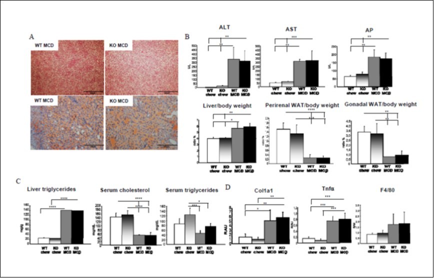
Download figure
Absence of PNPLA3 does not affect liver gene expression involved in de novo lipogenesis, fatty acid oxidation and uptake.
The sterol regulatory element binding protein 1c (SREBP1c) is a key regulator of PNPLA3 29 and regulates de novo lipogenesis, which accounts for 26% of the liver triglycerides in NAFLD 30. Therefore, we measured SREBP1c expression and its target genes stearoyl coA desaturase 1 (SCD-1) and fatty acid synthase (FASN) in MCD-fed WT and Pnpla3 KO mice. Generally, MCD diet reduced the expression of SREBP1c, SCD-1 and FASN in WT and Pnpla3 KO (Figure 2A). Interestingly, FASN and SCD-1 expressions were statistically reduced in Pnpla3 KO mice fed with MCD but not in WT (Figure 2A). Peroxisome proliferator activated receptor alpha (PPARα) is a nuclear receptor that senses fatty acids (FA) and directs them to b and w oxidation 31. PPARα expression was reduced by MCD diet in WT but not in Pnpla3 KO mice (Figure 2A), while PPARα expression was lowered at baseline in Pnpla3 KO compared to WT fed with chow. In line, the expression of the PPARα target gene carnitine palmitoyltransferase 1 alpha (CPT1α), involved in long chain fatty acid oxidation in mitochondria, was reduced in Pnpla3 KO compared to WT fed with chow diet, whereas MCD feeding did not influence its expression (Figure 2B). Another target of PPARα, the cytochrome P450 omega hydroxylase A14 (CYP4A14) catalyzes omega oxidation of medium chain FA and arachidonic acid and its liver deletion attenuates steatosis and fibrosis 32. CYP4A14 expression was reduced in WT mice upon MCD feeding, whereas Pnpla3 deletion did not affect its expression (Figure 2B). The peroxisomal acyl CoA oxidase (AOX) is the first enzyme involved in FA oxidation and a PPARα target 33. AOX gene expression was repressed by MCD feeding in WT and Pnpla3 KO (Figure 2B), while WT mice on chow diet had significantly higher AOX expression than Pnpla3 KO mice (Figure 2B). The fatty acid transporter 5 (FATP5/SLC27A5) is exclusively expressed in the liver and imports long chain fatty acids 34. MCD diet reduced FATP5 expression in a similar way in WT and Pnpla3 KO mice, respectively (Figure 2B). Moreover, FATP5 expression was lowered in KO mice under chow diet compared to WT (Figure 2B). The gene expression of the FA transporter CD36, a target of the peroxisome proliferator activated receptor gamma (PPARg), along with the expression of PPARg1 and PPARg2 were unchanged by MCD diet in WT and Pnpla3 KO mice (Figure 2C). PPARd, a nuclear receptor sensing non esterified FA obtained after lipolysis was similarly expressed upon MCD diet (Figure 2C). Finally, CYP7A1 expression, the rate limiting gene controlling bile acid synthesis, was unaffected by MCD in WT and Pnpla3 KO, in line with serum bile acids levels (Supplemental Fig1). Taken together, these results established that Pnpla3 deficiency did not influence FA synthesis, FA oxidation or bile acid synthesis in the MCD mouse model of steatohepatitis.
Figure 2. PNPLA3 does not affect liver gene expression involved in de novo lipogenesis, fatty acid oxidation and uptake. A: MCD diet reduced the expression of SREBP1c, SCD-1 and FASN in WT and Pnpla3 KO. FASN and SCD-1 expressions were statistically reduced in Pnpla3 KO mice fed with MCD but not in WT. PPARα expression was reduced by MCD diet in WT but not in KO mice, while its expression was lowered at baseline in KO compared to WT chow-fed. B: CPT1α expression was reduced in Pnpla3 KO compared to WT fed with chow diet. CYP4A14 expression was reduced in WT mice upon MCD feeding. AOX gene expression was repressed by MCD feeding in WT and Pnpla3 KO, while WT mice on chow diet had significantly higher AOX expression than KO. MCD diet reduced FATP5 expression in a similar way in WT and Pnpla3 KO, but FATP5 expression was lowered in KO under chow diet compared to WT. C: The gene expression of CD36, PPARg1, PPARg2 and PPARd remained unchanged by MCD diet in WT and Pnpla3 KO (n=5 per group). 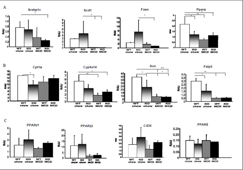
Download figure
Pnpla3/Adiponutrin deficiency does not affect liver injury, biliary fibrosis or damage in a BDL mouse model.
Next, we investigated whether Pnpla3/Adiponutrin deficiency affects mice subjected to bile duct ligation as a model for cholestasis (n= 3 per group) (Supplemental Fig.2). H&E staining and liver enzyme measurements in serum did not show any difference between WT- and Pnpla3-KO mice (Figure 3A, 3B&3C), despite a strong increase in ALT, AST, AP and bile acids levels in BDL-subjected mice irrespective of the genotype (Figure 3B&C). Sirius red staining (Figure 3A) and Col1a1 (Figure 3D) gene expression and protein levels (Supplemental Fig2) were comparable in WT and Pnpla3-KO mice upon BDL. In line, CK19 staining and gene expression (Figure 3A&3D) were unchanged between the genotypes. Moreover, inflammatory genes such as interleukin 6, Vcam1, desmin and Mcp1 had unchanged gene expression as shown by real time QPCR (Supplemental Fig2). Altogether, these data suggest that PNPLA3 deletion does not interfere with liver injury, fibrosis or cholangiocyte proliferation, inflammation and damage after BDL.
Figure 3. Pnpla3 deficiency does not affect liver injury, biliary fibrosis or damage in a BDL mouse model. A: H&E, Sirius red and CK19 staining did not show any differences between WT- and Pnpla3-KO BDL mice. B&C: ALT, AST, AP and bile acids levels increased in BDL-subjected mice irrespective of the genotype. D: Col1a1 and CK19 gene expression were unaffected by BDL between the genotypes (n=3 per group). 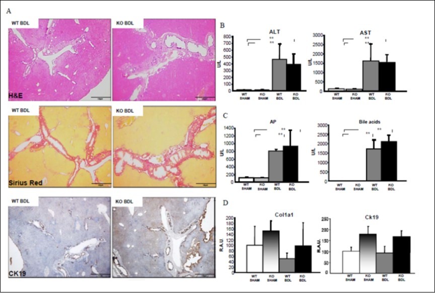
Download figure
Pnpla3/Adiponutrin deficiency does not affect biliary fibrosis or cholangitis in a DDC mouse model diet.
Male PSC patients encoding the I148M PNPLA3 variant may be at risk for reduced survival upon severe disease progression and bile duct stenosis requiring intervention who have 35. Therefore, and to test whether the absence of PNPLA3 influences bile duct injury and biliary fibrosis, WT and Pnpla3 KO mice (n= 5 per group) were fed with 0.1% of the xenobiotic DDC in normal chow diet for 12 days to induce sclerosing cholangitis and biliary fibrosis (Supplemental Fig.3) 24. H&E and cytokeratin 19 (CK19) staining showed similar bile duct injury and increased ductular reaction between WT and Pnpla3 KO (Figure 4A). Real time QPCR also demonstrated a similar increase in CK19 expression in WT and Pnpla3 KO (Figure 4B). In line, the liver enzymes ALT, AST and AP as well as serum bile acid levels increased upon DDC feeding but to the same degree in WT and Pnpla3 KO (Figure 4B). Liver weight to body weight as well as kidney weight to body weight ratios were unchanged between genotypes (Supplemental Fig3). Serum total cholesterol increased, when serum triglycerides levels decreased upon DDC feeding in a genotype independent manner (Supplemental Fig 3). Sirius Red staining showed a similar increase in fibrosis in WT and Pnpla3 KO mice (Figure 4A). In line, collagen 1a1 (Col1a1) expression measured by real time QPCR demonstrated the same gene induction in DDC-fed WT and KO (Figure 4B). Furthermore, spleen weight to body weight ratio decreased in DDC fed WT and KO to similar extent (Supplemental Fig3). We then studied the inflammatory response in the DDC model. F4/80 expression by real time QPCR was unchanged, whereas CD11b expression increased upon DDC in WT and Pnpla3 KO to the same degree (Figure 5). TNFa was only mildly increased in WT mice fed with DDC whereas Vcam-1, Timp1 and osteopontin expression was similarly increased by DDC feeding in WT and Pnpla3 KO (Figure 5). Taken together our data show that PNPLA3 deficiency does not affect ductular proliferation, inflammation or fibrosis in the DDC model.
Figure 4. Pnpla3 deficiency does not affect biliary fibrosis or cholangitis in a DDC mouse model diet. A: H&E, CK19 and Sirius red staining showed similar bile duct injury and increased ductular reaction between WT and Pnpla3 KO. Fig4B: ALT, AST, AP and serum bile acid levels increased upon DDC feeding to the same degree in WT and Pnpla3 KO. CK19 gene expression increased significantly between WT-chow and WT-DDC fed mice but not in Pnpla3 KO. Col1a1 had similar gene induction in DDC-fed WT and KO (n=5). 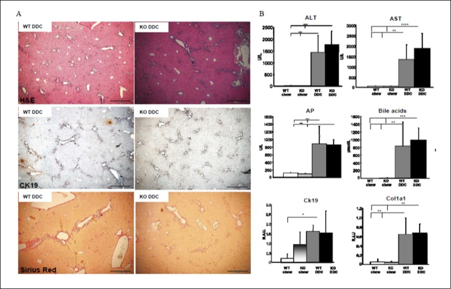
Download figure
Figure 5. Pnpla3 deficiency does not affect inflammatory parameters associated with biliary fibrosis or cholangitis in a DDC mouse model diet. F4/80 expression was unchanged, whereas CD11b expression increased upon DDC in WT and Pnpla3 KO to the same degree. TNFa expression increased in WT mice fed with DDC, when Vcam-1, Timp1 and osteopontin expression increased by DDC feeding to the same level in WT and Pnpla3 KO. 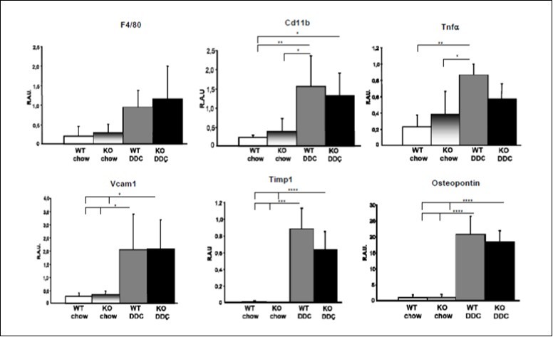
Download figure
PNPLA3 does not mitigate nor worsen CCl4 induced inflammation and fibrosis.
Carbon tetrachloride (CCl4) intoxication in rodents is a commonly used model of both acute and chronic liver fibrosis 36. To explore if lack of PNPLA3 alters the hepatic fibrotic response to chronic CCl4 intoxication, we injected WT and Pnpla3 KO mice (n= 6 per group) with CCl4 (2 ml/kg body weight of 1:4 diluted CCl4) (total injection volume 240 µl) twice a week for 4 weeks (Supplemental Fig.4). Control groups were treated with vehicle (100% olive oil, 2 ml/kg). First, H&E staining revealed a profound increase in fibrotic areas in the liver of both WT and Pnpla3 KO (Figure 6). Accordingly, the serum levels of liver enzyme ALT, but not of AST and AP, increased similarly after CCl4 injection in WT and KO (Figure 6). Serum bile acid levels also increased after CCl4 treatment but significantly more in Pnpla3 KO (Figure 6). Liver to body weight ratio increased significantly in Pnpla3 KO only after CCl4, while spleen to body weight ratio was unchanged and kidney to body weight ratio increased after CCl4 injection only in Pnpla3 KO compared to WT (Figure 6). Importantly, both Sirius Red staining and hydroxyproline determination, revealed that WT and Pnpla3 KO mice displayed similar degrees of fibrosis upon CCl4 injection (Figure 7). In line, real time QPCR measurement of collagen 1a1 and 1a2 showed a more pronounced increase of Col1a1 in Pnpla3KO, while conversely Col1a2 was reduced (Figure 7). In addition, alpha-SMA quantification by real time QPCR and staining desmin and MMP2 gene expression were also unchanged irrespective of the genotype. Finally, Timp1 expression increased to the same level in WT and Pnpla3 KO (Supplemental Fig4). Together our data suggest a role of Pnpla3 deficiency in fibrosis development in mice.
Figure 6. PNPLA3 does not mitigate nor worsen CCl4 induced inflammation and fibrosis. Fig6A. H&E staining revealed an increase in fibrotic areas in the liver of both WT and Pnpla3 KO. Fig6B: Serum levels of ALT, but not of AST and AP, increased similarly after CCl4 injection in WT and KO. Serum bile acids levels significantly increased after CCl4 treatment in Pnpla3 KO. Fig6C: Liver to body weight ratio increased significantly in Pnpla3 KO only after CCl4, while spleen to body weight ratio was unchanged and kidney to body weight ratio increased after CCl4 injection only compared to WT control (n=6 per group). 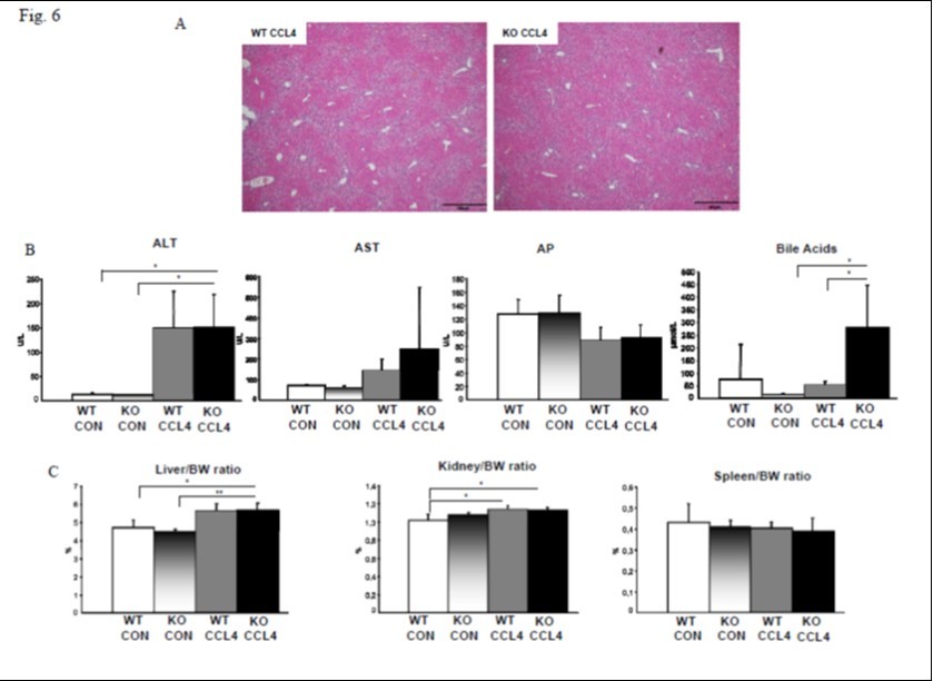
Download figure
Figure 7. PNPLA3 does not mitigate nor worsen CCl4 induced hydroxyproline and collagen gene expression. Fig7A&B. Sirius Red staining and hydroxyproline measurement showed that WT and Pnpla3 KO mice had similar degrees of fibrosis upon CCl4 injection. Fig7C: Col1a1 expression increased more in Pnpla3KO upon CCl4, when Col1a2 expression was reduced compared to WT mice. 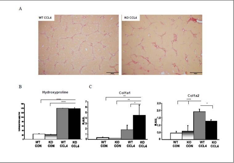
Download figure
Discussion
Previous studies have demonstrated that mice lacking Pnpla3 do not show differences in the development of steatosis when challenged by chow diet, fasting/refeeding, LXR agonists, high sucrose, high fat diet, or bred with leptin deficient mice 22, 37. Knock-in models containing the I148M human mutation inserted into the Pnpla3 mouse locus required extreme dietary challenges with high fat, high cholesterol and fructose for 25 38 to 52 weeks 39 to obtain a liver disease and the addition of ethanol to achieve HCC development in 25 weeks 40. This lack of metabolic phenotype in the Pnpla3 deficient mouse and the requirement of long dietary challenges in the knock-in model was puzzling when compared to the human data showing a key role for the loss of function gene variant in the pathogenesis of NAFLD 13 and progression of liver diseases irrespective of their aetiologies 1, 2, 3, 4, 5. Therefore, our study aimed to address whether Pnpla3 deficiency in mice interferes with the degree of inflammation and fibrosis in models of more pronounced liver injury. Our data revealed that lack of Pnpla3 in mice does not further impact the development of steatohepatitis or fibrosis induced by MCD diet. Moreover, minor signaling differences in the PPARα pathway were only found under chow diet feeding between WT and Pnpla3 KO mice. Bile duct ligation and DDC feeding as model for sclerosing cholangitis and biliary fibrosis did not reveal any differences in cholestatic injury and biliary fibrosis between WT and Pnpla3 KO mice. Finally, fibrosis induced by CCl4 as more general fibrosis model also did not show phenotypic differences in Pnpla3 KO mice compared to WT.
PNPLA3 was first identified as a gene involved in white adipocyte differentiation in vitro 6. PNPLA3 is the closest homolog of PNPLA2, also known as adipose triglyceride lipase (ATGL), which plays a key role in NAFLD development 28. These enzymes share a similar structure with three trans-membrane domains in the N-terminus and a fourth in the C-terminus, however their catalytic site with a dyad Ser-Asp is different from other lipases 41. Remarkably, human and mouse PNPLA3 expression pattern diverge significantly with the human gene being highly expressed in liver, retina, kidney, white adipose tissue and brain, while the mouse gene is mostly expressed in white adipose tissue and only to low extent in hepatocytes and HSC 6, 8. In addition, when human hepatocytes are all expressing PNPLA3 at high rate, only a fraction of mouse hepatocytes do so 8, 42. Despite these gene expression differences and a relatively low homology between the mouse and human protein with only 53% of amino-acids conservation 6, human and mouse proteins showed a similar structure especially around the catalytic site using Alphafold 43. The I148M polymorphism in PNPLA3 was identified as a key factor in multiple liver etiologies, conferring risk for NAFLD development, inflammation 13, 44, in HCC progression 45 and in fibrosis progression after liver transplantation in hepatitis C 46, but also in hepatic insulin resistance 47. Since this polymorphism is located in the vicinity of the active catalytic site, it could affect the enzyme activity. Several studies generated conflicting results about the role of PNPLA3 being an acyl transferase 10, or a retinol hydrolase in HSC 17, or affecting VLDL secretion in human hepatocytes in vitro 48. Thus, the real biochemical function of PNPLA3 is still unclear and whether the I148M mutation is a gain or loss of function is also still far from evident. At the molecular level PNPLA3 regulation is under the control of SREBP1c and LXR signaling, two transcription factors involved in de novo lipogenesis, therefore pointing toward a role for PNPLA3 in lipid catabolism 8. It is however important to consider that lipogenesis differs substantially between species, while human lipogenesis takes place mainly in liver, mouse lipogenesis is more adipocyte mediated 49, 50 and hence the expression levels of SREBP1c and LXR and PNPLA3 8, 51. Recently, in human HSC our group showed that PNPLA3 I148M mutation resulted in dysregulated LXR and PPARg signaling due to abnormal phosphorylation and accumulation of cholesterol leading to exacerbated pro-fibrogenic and inflammatory response 18, 19.
PNPLA3 I148M was found to accumulate at lipid droplets 14 and evade ubiquitylation 15, thereby disrupting triglyceride hydrolysis due to a competition between PNPLA3 I148M and CGI-58 as main cofactor required by PNPLA2/ATGL to hydrolyze triglycerides from the lipid droplet 52, 16. ATGL activity on lipid droplets leads to the release of linoleic acid, an essential fatty acid and PPARα ligand in heart 53 and liver 28. Therefore, we might expect that Pnpla3 knock-out mice by allowing a stable Cgi-58 interaction with Atgl, would increase Atgl activity and linoleic acid release thereby stimulating PPARα and its target gene expression. However, our data revealed that PPARα expression and its target genes Aox and Cpt1α were only reduced in Pnpla3 KO mice fed with a chow diet, an effect lost under MCD, DDC or CCl4 challenge. (Figure 2). PPARα KO mice displayed a fatty liver phenotype at baseline and do not respond to synthetic agonists fibrates 33 and have disrupted fatty acid oxidation 54. As such the reduction of PPARα signaling and key genes involved in fatty acid oxidation in Pnpla3 KO could at least in part contribute to the key role of PNPLA3 in fatty liver development. However, Pnpla3 KO mice did not develop more severe steatohepatitis upon MCD challenge. This apparent contradiction, together with the conflicting results obtained between human and mouse, could be due to the fact that humans express very low levels of PPARα compared to mouse 55 and as such a 20% reduction in PPARα signaling in mouse would not be expected to translate into pathophysiological consequences. Therefore, insights from mouse models over-expressing the PNPLA3 I148M variant and the WT protein at similar rates obtained under the control of the apoE promoter 56 are of interest. These mice did not display changes either in PPARα expression or in its target genes Cpt1α and Aox, 56 thus questioning the existence of a PNPLA3/ATGL/CGI-58 regulatory loop in mice. It is thus tempting to speculate that mouse and human PNPLA3 having different expression patterns and not displaying the same polymorphisms evolved to adapt to different stress conditions and have different roles, which are not redundant. Due to all these differences the PNPLA3 KO mouse model is limited in terms of mimicking the human situation, a problem exacerbated by the short time of the challenges applied and the extreme metabolic turn-over seen in mice.
Conclusion
Overall, the limitations of mouse models studying the role of PNPLA3 variants seem to require extreme dietary challenges in knock-in models for a long time period, or in the case of chimeric mice, having human hepatocytes integrated in their livers, the use of mouse allowing human cells engraftment lacking B, T and NK cells 57. Since the expression pattern of PNPLA3 is different between species 51 and is controlled in a species-specific manner by different transcription factors, it seems that knocking in the human PNPLA3 gene with its entire regulating sequences would be a more relevant, yet ambitious model to unravel the role(s) of PNPLA3 in lipid metabolism and become a valid preclinical model.
Authors contributions
Robert McMahon and Thierry Claudel performed and analyzed all experiments, wrote the manuscript and made the figures, Claudia Fuchs and Robert McMahon performed the BDL experiment, Tatjana Stojakovic and Hubert Scharnagl performed serum parameters measurements, Michael Trauner supervised the work, revised the manuscript and provided funding; all authors critically revised the manuscript, provided critical intellectual input and approved the final version of the manuscript
Abbreviations
- ALT: alanine amino transferase
- AP: alkaline phosphatase
- AOX: acyl CoA oxidase
- AST: aspartate amino transferase
- BDL: bile duct ligation
- CCl4: carbon tetrachloride
- CK19: cytokeratin 19
- Col1a1: collagen 1a1
- CPT1α: carnitine palmitoyltransferase 1 alpha
- DDC: 3
-
5-diethoxycarbonyl-1
- FASN: fatty acid synthase
- FATP5: fatty acid transporter 5
- HCC: hepatocellular cancer
- H&E: haematoxylin and eosin
- HSC: hepatic stellate cells
- Il1β: interleukin 1 beta
- KC: Kupffer cells
- MASLD: metabolic dysfunction-associated steatotic liver disease
- MCD: methionine-choline deficient
- NAFLD: non-alcoholic fatty liver disease
- NASH: non-alcoholic steatohepatitis
- PNPLA3: patatin-like phospholipase domain-containing protein 3
- PPARα: peroxisome proliferator activated receptor alpha
- PPARg: peroxisome proliferator activated receptor gamma
- PSC: primary sclerosing cholangitis
- SCD-1: stearoyl coA desaturase 1
- SREBP1c: sterol regulatory element binding protein 1c
- TG: triglycerides
References
- 1.F V Bruschi, Tardelli M, Claudel T, Trauner M. (2017) PNPLA3 expression and its impact on the liver: current perspectives. Hepatic Medicine:. Evidence and Research 9, 55-66.
- 2.Linden D, Romeo S. (2023) Mini-Review for Journal of HepatologyTherapeutic opportunities for the treatment of NASH with genetically validated targets. J Hepatol
- 3.Cherubini A, Casirati E, Tomasi M, Valenti L. (2021) PNPLA3 as a therapeutic target for fatty liver disease: the evidence to date. Expert Opin Ther Targets 25, 1033-1043.
- 4.Tardelli M, F V Bruschi, Trauner M. (2020) . The Role of Metabolic Lipases in the Pathogenesis and Management of Liver Disease. Hepatology 72, 1117-1126.
- 5.J C Cohen, J D Horton, H. (2011) Human fatty liver disease: old questions and new insights. Science 332, 1519-23.
- 6.Baulande S, Lasnier F, Lucas M, Pairault J. (2001) Adiponutrin, a transmembrane protein corresponding to a novel dietary- and obesity-linked mRNA specifically expressed in the adipose lineage. J Biol Chem 276, 33336-44.
- 7.C M Jenkins, D J Mancuso, Yan W, H F Sims, Gibson B. (2004) Identification, cloning, expression, and purification of three novel human calcium-independent phospholipase A2 family members possessing triacylglycerol lipase and acylglycerol transacylase activities. J Biol Chem 279, 48968-48975.
- 8.Huang Y, He S, J Z Li, Y K Seo, T F Osborne. (2010) A feed-forward loop amplifies nutritional regulation of PNPLA3. Proc Natl Acad Sci U S A 107, 7892-7897.
- 9.He S, McPhaul C, J Z Li, Garuti R, Kinch L. (2010) A sequence variation (I148M) in PNPLA3 associated with nonalcoholic fatty liver disease disrupts triglyceride hydrolysis. J Biol Chem 285, 6706-15.
- 10.M Schoiwohl Kumari, Chitraju G, Paar C, Cornaciu M, I. (2012) Adiponutrin functions as a nutritionally regulated lysophosphatidic acid acyltransferase. Cell Metab 15, 691-702.
- 11.M E Rinella, J V Lazarus, Ratziu V, S M Francque, A J Sanyal. (2023) A multi-society Delphi consensus statement on new fatty liver disease nomenclature. J Hepatol
- 12.Valenti L, Dongiovanni P, Corradini Ginanni, Burza S, A M et al. (2013) PNPLA3 I148M variant and hepatocellular carcinoma: a common genetic variant for a rare disease. Dig Liver Dis 45, 619-24.
- 13.S Kozlitina Romeo, Xing J, Pertsemlidis C, Cox A, D. (2008) Genetic variation in PNPLA3 confers susceptibility to nonalcoholic fatty liver disease. Nat Genet 40, 1461-1465.
- 14.Smagris E, BasuRay S, Li J, Huang Y, K M Lai. (2015) Pnpla3I148M knockin mice accumulate PNPLA3 on lipid droplets and develop hepatic steatosis. Hepatology 61, 108-18.
- 15.BasuRay S, Smagris E, J C Cohen, H. (2017) The PNPLA3 variant associated with fatty liver disease (I148M) accumulates on lipid droplets by evading ubiquitylation. Hepatology 66, 1111-1124.
- 16.Wang Y, Kory N, J C Cohen, H. (2019) PNPLA3, CGI-58, and Inhibition of Hepatic Triglyceride Hydrolysis in Mice. Hepatology.
- 17.Pirazzi C, Valenti L, B M Motta, Pingitore P, Hedfalk K. (2014) PNPLA3 has retinyl-palmitate lipase activity in human hepatic stellate cells. Hum mol genet. 23, 4077-85.
- 18.F V Bruschi, Claudel T, Tardelli M, Caligiuri A, T M Stulnig. (2017) The PNPLA3 I148M variant modulates the fibrogenic phenotype of human hepatic stellate cells. Hepatology 65, 1875-1890.
- 19.F V Bruschi, Claudel T, Tardelli M, Starlinger P, Marra F. (2019) PNPLA3 I148M Variant Impairs Liver X Receptor Signaling and Cholesterol Homeostasis in Human Hepatic Stellate Cells. Hepatol Commun 3, 1191-1204.
- 20.F V Bruschi, Tardelli M, Einwallner E, Claudel T, Trauner M. (2020) PNPLA3 I148M Up-Regulates Hedgehog and Yap Signaling in Human Hepatic Stellate Cells. Int J Mol Sci 21.
- 21.Farkkila M, Kautiainen H, Tenca A, Jokelainen K, Arola J. (2022) PNPLA3 allele frequency has no impact on biliary bile acid composition or disease course in patients with primary sclerosing cholangitis. PLoS One 17, 0277084.
- 22.M K Basantani, M T Sitnick, Cai L, D S Brenner, N P Gardner. (2011) Pnpla3/Adiponutrin deficiency in mice does not contribute to fatty liver disease or metabolic syndrome. J Lipid Res 52, 318-29.
- 23.P Zollner Fickert, Fuchsbichler G, Stumtner A, Weiglein C, A H. (2002) Ursodeoxycholic acid aggravates bile infarcts in bile duct-ligated and Mdr2 knockout mice via disruption of cholangioles. Gastroenterology 123, 1238-1251.
- 24.Fickert P, Stoger U, Fuchsbichler A, Moustafa T, Schnabl B. (2007) A new xenobiotic-induced mouse model of sclerosing cholangitis and biliary fibrosis. Am J Pathol 171, 525-536.
- 25.Y H Paik, Iwaisako K, Seki E, Inokuchi S, Schnabl B. (2011) The nicotinamide adenine dinucleotide phosphate oxidase (NOX) homologues NOX1 and NOX2/gp91(phox) mediate hepatic fibrosis in mice. Hepatology 53, 1730-41.
- 26.Folch J, Lees M, Stanley Sloane, H G. (1957) A simple method for the isolation and purification of total lipides from animal tissues. J Biol Chem 226, 497-509.
- 27.Jha P, Knopf A, Koefeler H, Mueller M, Lackner C. (2014) Role of adipose tissue in methionine-choline-deficient model of non-alcoholic steatohepatitis (NASH). Biochimica et biophysica acta 1842. 959-70.
- 28.Jha P, Claudel T, Baghdasaryan A, Mueller M, Halilbasic E. (2014) Role of adipose triglyceride lipase (PNPLA2) in protection from hepatic inflammation in mouse models of steatohepatitis and endotoxemia. Hepatology 59, 858-69.
- 29.Dubuquoy C, Robichon C, Lasnier F, Langlois C, Dugail I. (2011) Distinct regulation of adiponutrin/PNPLA3 gene expression by the transcription factors ChREBP and SREBP1c in mouse and human hepatocytes. Journal of hepatology 55, 145-53.
- 30.K L Donnelly, C I Smith, S J, Jessurun J, Boldt. (2005) Sources of fatty acids stored in liver and secreted via lipoproteins in patients with nonalcoholic fatty liver disease. J Clin invest 115, 1343-1351.
- 31.Wang Y, Nakajima T, F J Gonzalez, Tanaka N. (2020) PPARs as Metabolic Regulators. in the Liver: Lessons from Liver-Specific PPAR-Null Mice. International journal of molecular sciences 21.
- 32.Zhang X, Li S, Zhou Y, Su W, Ruan X. (2017) Ablation of cytochrome P450 omega-hydroxylase 4A14 gene attenuates hepatic steatosis and fibrosis. Proceedings of the National Academy of Sciences of the United States of America 114 3181-3185.
- 33.S, Pineau T, Drago J, E J Lee, J W Owens. (1995) Targeted disruption of the alpha isoform of the peroxisome proliferator-activated receptor gene in mice results in abolishment of the pleiotropic effects of peroxisome proliferators. Mol Cell Biol 15, 3012-3022.
- 34.H Baillie Doege, R A Ortegon, A M Tsang, Wu B, Q. (2006) Targeted deletion of FATP5 reveals multiple functions in liver metabolism: alterations in hepatic lipid homeostasis. Gastroenterology 130, 1245-58.
- 35.Friedrich K, Rupp C, J R Hov, Steinbrunner N, K H Weiss. (2013) A frequent PNPLA3 variant is a sex specific disease modifier in PSC patients with bile duct stenosis. PLoS ONE 8, 58734.
- 36.Tamayo Perez, R. (1983) Is cirrhosis of the liver experimentally produced by CCl4 and adequate model of human cirrhosis?. Hepatology 3, 112-20.
- 37.Chen W, Chang B, Li L, Chan L. (2010) Patatin-like phospholipase domain-containing 3/adiponutrin deficiency in mice is not associated with fatty liver disease. Hepatology 52, 1134-42.
- 38.Linden D, Ahnmark A, Pingitore P, Ciociola E, Ahlstedt I. (2019) Pnpla3 silencing with antisense oligonucleotides ameliorates nonalcoholic steatohepatitis and fibrosis in Pnpla3 I148M knock-in mice. Mol Metab 22, 49-61.
- 39.Su H, Haque M, Becker S, Edlund K, Duda J. (2023) Long-term hypercaloric diet exacerbates metabolic liver disease in PNPLA3 I148M animals. Liver Int.
- 40.Patsenker E, V R Thangapandi, Knittelfelder O, Palladini A, Hefti M. (2022) The PNPLA3 variant I148M reveals protective effects toward hepatocellular carcinoma in mice via restoration of omega-3 polyunsaturated fats. J Nutr Biochem 108, 109081.
- 41.P A Wilson, S D Gardner, N M Lambie, S A Commans, D J Crowther. (2006) Characterization of the human patatin-like phospholipase family. Journal of lipid research 47, 1940-9.
- 42.Hoekstra M, Li Z, J K Kruijt, M Van Eck, Berkel T J Van. (2010) The expression level of non-alcoholic fatty liver disease-related gene PNPLA3 in hepatocytes is highly influenced by hepatic lipid status. J Hepatol 52, 244-51.
- 43.Ramandi A, A M Diehl, A J Sanyal, Jong Y P de. (2025) . Experimental Models to Investigate PNPLA3 in Liver Steatosis. Liver Int 45 70091.
- 44.E K Speliotes, J L Butler, C D Palmer, B F Voight, J N Hirschhorn. (2010) PNPLA3 variants specifically confer increased risk for histologic nonalcoholic fatty liver disease but not metabolic disease. Hepatology 52, 904-912.
- 45.Y L Liu, G L Patman, J B Leathart, A C Piguet, A D Burt. (2014) Carriage of the PNPLA3 rs738409 C >G polymorphism confers an increased risk of non-alcoholic fatty liver disease associated hepatocellular carcinoma. J Hepatol 61, 75-81.
- 46.Dunn W, Neil O, Zhao M, Wu J, C H Roberts et al. (2014) Donor PNPLA3 rs738409 genotype affects fibrosis progression in liver transplantation for hepatitis C. Hepatology 59, 453-60.
- 47.O P Zaharia, Strassburger K, Knebel B, Kupriyanova Y, Karusheva Y. (2020) . Role of Patatin-Like Phospholipase Domain-Containing 3 Gene for Hepatic Lipid Content and Insulin Resistance in Diabetes. Diabetes Care 43, 2161-2168.
- 48.Pirazzi C, Adiels M, M A Burza, R M Mancina, Levin M. (2012) Patatin-like phospholipase domain-containing 3 (PNPLA3) I148M (rs738409) affects hepatic VLDL secretion in humans and in vitro. Journal of Hepatology 57, 1276-1282.
- 49.Diraison F, Yankah V, Letexier D, Dusserre E, Jones P. (2003) Differences in the regulation of adipose tissue and liver lipogenesis by carbohydrates in humans. J Lipid Res 44, 846-53.
- 50.A K Stoeckman, Ma L, H C Towle. (2004) Mlx is the functional heteromeric partner of the carbohydrate response element-binding protein in glucose regulation of lipogenic enzyme genes. J Biol Chem 279, 15662-9.
- 51.A C Lake, Sun Y, J L Li, J E Kim, J W. (2005) Expression, regulation, and triglyceride hydrolase activity of Adiponutrin family members. J Lipid Res 46, 2477-2487.
- 52.BasuRay S, Wang Y, Smagris E, J C Cohen, H. (2019) Accumulation of PNPLA3 on lipid droplets is the basis of associated hepatic steatosis. Proc Natl Acad Sci U S A 116, 9521-9526.
- 53.Haemmerle G, Moustafa T, Woelkart G, Buttner S, Schmidt A. (2011) ATGL-mediated fat catabolism regulates cardiac mitochondrial function via PPAR-alpha and PGC-1. Nature medicine 17, 1076-85.
- 54.Aoyama T, J M Peters, Iritani N, Nakajima T, Furihata K. (1998) Altered constitutive expression of fatty acid-metabolizing enzymes in mice lacking the peroxisome proliferator-activated receptor alpha (PPARalpha). J Biol Chem 273, 5678-5684.
- 55.C N Palmer, M H Hsu, K J Griffin, J L Raucy, E F Johnson. (1998) Peroxisome proliferator activated receptor-alpha expression in human liver. Mol Pharmacol 53, 14-22.